Sunday, October 3, 2010
Recent CT scan results
IMPRESSION:
1. Findings consistent with cirrhosis. No evidence of cellular carcinoma. Patchy hypodensity in the right lobe of liver and a more focal subtle lesion with mild mass effect lateral segment left lobe liver measuring 3.1 cm in diameter, more conspicuous and slightly larger compared to 6-16-10 when it measured 2.8 centimeters in diameter. Recommend correlation with ultrasound and dynamic MR.
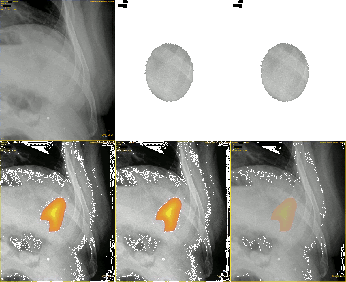
2. Slowly enlarging lytic lesion of the anterolateral left seventh rib. Considerations include fibrous dysplasia, aneurysmal bone cyst, myeloma and metastasis. Recommend further evaluation
Kind of scary. Metastasis?!?!?!?! WTF!!!!!
Subscribe to:
Post Comments (Atom)




















No comments:
Post a Comment