
Here is a pretty good article about diagnosing Hepato Cellular Carcinoma, also known as LIVER CANCER, or Primary Liver Cancer, as apposed to secondary Liver Cancer aka liver metastases.
It seems to this liver that the techneque to diagnose these CELLS GONE WILD is as much art as it is science, and even maybe a small dose of VOODOO thrown in for good/bad measure.
Any way the LUMP being hunted and stalked by the hepatology department at CU, headed by the World Famous Hepatologist Jane Berman (name changed to protect the innocent)
is still at large, not being called an HCC and not seen clearly due to technical matters, which are detailed in the linked article. So, if you are a friend or daughter, or mother/dad, or sister or brother of the same human family as billybob belongs to, and you are curious what the hairy hell Bobby is talking about when the subject comes up, just check out the part on "washout" as far as the CT scan is concerned... Think about why Sawyer's Mommy doesn't drink from his bottle and employs the super cool bottle cap trick, to avoid washout... Liver nodules which have scar tissue, can flood with the blood which is not dyed by iodine, mixing it with dyed blood, at the precise moment they are supposed to light up like the Vegas strip, because of an auto injection of radioactive iodine, they get "washout", and they hide from the camera. It seems that this is the only time during a CT scan this could occur in the whole human body. Scarred livers have blood flowing in all kinds of crazy directions. Dual phase CT scan is supposed to take pictures of non radio active tissue and then it takes pictures of the same tissue a moment later with a shot of that iodine, to light it up all the blood vessels... but in order to see a small just forming tumor, it has to be snapped at just the right moment, and the body rides through the scanner for several seconds, moving, and by the time the camera catches the part where the suspected tumor is located, the dye may have "washed out". Portal hypertension in action. Backward blood flow.
Here is what the radiologist said about it-copied in shortened version:
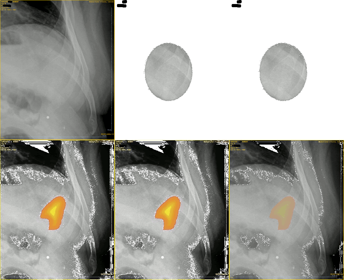
The vague hypodensity bulging the capsule in the left lateral segment anteriorly, best seen on series 9 image #38 is not significantly changed. No lesion here is more conspicuous on delayed imaging suggesting possible early washout. Additionally, there is vague mottled hypodensity throughout the right lobe of the liver.
IMPRESSION:
1. No change in subtle left lobe of the liver lateral segment anterior hypodensity which may have early washout. Diagnostic considerations include regenerative nodules after infarct, focal fatty infiltration, although mass cannot be excluded, given recent normal ultrasound and stability, this is felt to be less likely.
What else is there to say, lol.













1 comment:
Uh, so they didn't get a good picture of Billy Bob? Please have Billy Bob's human host call his daughter :)
Post a Comment